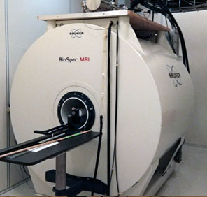
7T Bruker Scanner
 |
Location: Elliman Building Room 0121 421 E. Canfield Detroit, MI 48201 Contact: Robin Roberts B.S. ph: 313-577-1747 |
Submit New Proposal Book Scan Time |
The Bruker BioSpec® 70/30 USR horizontal bore MRI system is designed for performing preclinical and molecular MRI. The high field MR scanner can perform both magnetic resonance imaging (MRI) and magnetic resonance spectroscopy (MRS) for preclinical, pharmaceutical, and fundamental research. The scanner is actively-shielded 7T magnet with a 30-cm bore and cryo-refrigeration that minimizes the time and cost associated with magnet maintenance. The system is equipped with a 12 cm gradient and shim coil set (B-GA12S HP), capable of generating maximum gradient amplitude of 660 mT/m, rise time of 80 to 120 us, slew rate of 4570 T/m/s, and 4-channel receiver for multi-coil operation.
The operating console has ParaVision 360 (v2.0pl1) acquisition workplace, which features the following: built-in parallel acquisition; push-button GRAPPA reconstruction; EPI, navigator techniques for motion reduction; ultra-short TE acquisition; Half-Fourier encoding; self-gated IntraGate; real-time display of acquired and reconstructed data; sophisticated data archiving including DICOM export; and enhanced 2D and 3D data visualization.
The MRI suite is equipped with a dedicated isoflurane anesthesia system, temperature controlled platform, cardiac gating, and respiratory gating. This 30 cm bore system achieves sub-millimeter spatial resolution with variable, user-selectable, field-of-view and sensitivity. The acquisition times range from <1 s to 30 min depending on chosen resolution, field-of-view and function.
Services Provided:
- MR Protocol development and application
- Research Collaboration and Consultation
- Mentoring and Training
Equipment Features:
- High end UltraShield™ Refrigerated (USR) magnet technology.
- Helium zero-boil-off and Nitrogen free magnet technology for reduced maintenance costs and longer service intervals.
- Scalable AVANCE NEO RF architecture incorporates up to 16 receiver and 6 transmitter channels.
- Parallel imaging (GRAPPA) for almost all applications including EPI.
- Multiple transmit imaging applications.
- High performance BGA-S gradients with highest amplitudes and slew rates, shim strengths, and duty cycles optimized for high field MRI.
- IntraGate™ - Self gated steady-state cardiac imaging (no external sensor hardware and triggering devices).
- Phased-array RF coil technology for maximum sensitivity and minimum scan times.
- ParaVision® - Intuitive user interface, for multi-dimensional MRI/MRS data acquisition, reconstruction, analysis and visualization.
Gradient/Shim
1H Transmit and Receive Channel
The electronics cabinet is equipped for digital signal generation and detection of 1H MRI/MRS experiments. The following RF coil configurations are supported:- Transmit and receive volume or surface coil
- Combination of transmit volume coil with receive-only surface coil
- Combination of transmit volume coil with receive-only array coil
RF Amplifier for Proton MRI/MRS excitation
- One 1 kW transmitter for 1H excitation
RF Quadrature Combiner
- Active quadrature combiner for a maximal input power of 1kW.
RF Tune/Match Adapter
- The tune and match adapter provides tune and match capabilities for circularly polarized MRI coils without integrated preamplifier
RF Interface Adapter
The RF interface adapter allows the operation of RF coils that are not equipped with standard Bruker RF connector e.g. home built or third party RF coils. The adapter supports the following socket types:
- N-type connector for 1H, X high power transmit/receive coils
- TWINAX connector providing the pin-diode voltage for active coil decoupling
- BNC-type connector for 1H, X low power transmit/receive coils
- 14 pin DIN-type connector for receive only surface coils
- 4 RF pin / 14 DC pin connector for 4 channel receive only array coils
High Power Gradient Amplifier Upgrade
- A complete 3 channel IECO MRI gradient amplifier cabinet providing 300 A / 500 V output power.
Shim Amplifier
Shim amplifier (6 channels, max current 10 A)ParaVision Acquisition Workplace
The ParaVision 360 acquisition workplace is delivered as part of the MR instrument and includes the following items and functionality:
Computer hardware:
- 64 bit Linux multicore workstation, 64 GB RAM, 3TB hard disk
- Full HD monitor, keyboard and mouse
- Database browser
- Network data transfer between multiple ParaVision workplaces
- Archiving on DVD and data server
- Image Analysis and Reporting: ROI, Profiles, Image Sequence Analysis, Image Algebra, Report generation
Standard Imaging Sequences and protocols:
The Study Planning package includes the following methods and protocols:
- Fast gradient echo (FLASH)
- Multiple slice multiple spin echo (MSME)
- Fast spin echo (RARE)
- T1-weighted fast spin echo (MDEFT)
- Steady state free precision imaging (FISP)
- Non-localized pulse & acquire (SINGLEPULSE, NSPECT)
- Dixon fat/water imaging
- CEST imaging
- Susceptibility weighted Imaging (SWI)
- Automatic B0 field map shimming method
- Automatic B1 field calibration (B1MAP)
- Triggered acquisition
Parallel Receiver Upgrade: 1 → 4 Channels
A fully equipped parallel proton-receiver configuration allows the application of 1H parallel imaging methods. The upgrade comprises:
- 3 additional 1H receiver channels for a total of 4 channels
- GRAPPA based parallel imaging reconstruction
3D Visualization and Analysis
A3D data visualization and analysis extension package enhances the basic viewing functionality of ParaVision by:
- 3D interactive viewing
- Maximum intensity projection (MIP)
- Multi-planar secondary image reconstruction (MPR)
- Surface rendering
- Movie generation
Multimodal Data Import and Export
The multimodal data functionality extension package of ParaVision allows to import and export DICOM data of the following imaging modalities:
- Magnetic Resonance Imaging (MRI)
- Magnetic Particle Imaging (MPI)
- Magnetic Resonance Imaging (MRI)
- Magnetic Particle Imaging (MPI)
Cardio Package
The cardio application package of ParaVision includes all methods and ParaVision functionality tools required for triggered or self-gated cardiac MRI.
Methods and protocols:
- Flow-compensated fast gradient echo imaging (FcFLASH)
- Self-gated cardiac imaging (IntraGate_FLASH)
ParaVision functionality extension:
The software feature of the processing workplace is extended by:
- Self-gated cardiac image reconstruction
- Self-gated cardiac imaging (IntraGate_UTE)
Diffusion Package
The diffusion application package of ParaVision includes all methods and ParaVision functionality tools required for diffusion weighted or diffusion tensor MRI.
Methods and protocols:
- Spin echo based diffusion tensor imaging (DTI_STANDARD)
- Echo planar based diffusion tensor imaging (DTI_EPI)
- Spiral based diffusion tensor imaging (DTI_SPIRAL)
ParaVision functionality extension:
The software feature of the processing workplace is extended by:
- DTI analysis and visualization
Angiography Package
The angiography application package of ParaVision includes all methods and ParaVision functionality tools required for angiography and velocity MRI.
Methods and protocols:
- Flow-compensated fast gradient echo imaging (FcFLASH)
- Phase contrast angiography and quantitative flow mapping (FLOW_MAP)
fMRI and DCE Package
The functional MRI and the dynamic contrast enhanced (DCE) application package of ParaVision includes all methods and ParaVision functionality tools required for functional and dynamic contrast enhanced kinematic MRI.
Methods and protocols:
- Spin echo based echo planar imaging (EPI)
- Gradient echo based echo planar imaging (EPI)
- Flow-sensitive alternating inversion recovery (FAIR_RARE, FAIR_EPI)
ParaVision functionality extension:
- Functional Image Analysis Tool
Short Echo Time Package
The short echo time application package of ParaVision includes all methods required for imaging samples with short echo times by means of MRI.
Methods and protocols:
- Fast spin-echo imaging with very short echo times (RAREST)
- Spiral imaging (SPIRAL)
- Ultra short echo time imaging (UTE, UTE3D)
- Zero echo time 3D imaging (ZTE)
Relaxation Package
The relaxation application package of ParaVision includes all methods required for T1/T2 based MRI contrasts and MR relaxometry.
Methods and protocols:
- Multi gradient echo imaging (MGE)
- Variable repetition time fast spin echo (RAREVTR)
- T1 echo planar imaging (T1_EPI)
- T2* echo planar imaging (T2S_EPI)
- T2 echo planar imaging (T2_EPI)
Spectroscopy Package
The spectroscopy application package of ParaVision includes all methods and ParaVision functionality tools required for spatially resolved MRS.
Methods and protocols:
- Spectroscopic imaging (CSI)
- Echo planar spectroscopic imaging (EPSI)
- Localized single voxel spectroscopy (PRESS, STEAM, ISIS)
- High-resolution NMR pulse sequence library
ParaVision functionality extension:
- Spectral acquisition, processing and analysis software (TopSpin)
Transmit/Receive Volume Coil for Mice and Rats MT- 72 mm
This volume coil is optimized for cross coil setups, i.e. for use as transmit RF coil in combination with receive-only surface coils and arrays. The coil can also be used as receive volume coil. Small rodents up to 450 g body weight can be examined. Designed for use in B-GA9S or bigger gradient coils.
- 1H transmit and receive capabilities with active coil detuning
- Circularly
Rat Body Volume Coil - 72 mm
Volume RF coil designed for rodent body investigations up 450 g body weight. Designed for use in B-GA12S or bigger gradient coils.
- 1H transmit and receive capabilities
- Circularly polarized
- Outer/inner diameter 112 mm / 72 mm
Mouse Body Volume Coil - 35 mm
Volume RF coil for mouse body investigations. Designed for use in B-GA6S or bigger gradient coils.
- 1H transmit and receive capabilities
- Circularly polarized
- Outer/inner diameter 59 mm / 35 mm
Rat Brain Surface Coil
Anatomically shaped rat brain surface coil.
- 1H receive-only capabilities with active coil detuning
- Circularly polarized
- Integrated preamplifiers
Rat Brain Array Coil
Anatomically shaped 4 elements rat brain surface coil.
- 1H receive-only capabilities with active coil detuning
- 2x2 coil elements topology
- Integrated preamplifiers
Mouse Brain Array Coil – 4 Channels
Anatomically shaped four element array coil for mouse brain investigations.
- 1H receive-only capabilities with active coil detuning
- 2x2 coil elements topology
- Integrated preamplifiers
Low Noise Preamplifier for Planar Surface Coils
Low noise preamplifier to be used with the planar surface coils listed below. The preamplifier is to be mounted on a split cradle. Designed for use in B-GA9S or bigger gradient coils.
Multi-Purpose Planar Surface Coil – 10 mm
1H receive-only surface coil
- 1H receive-only capabilities with active coil detuning
- Single loop coil with flexible cable
- Diameter 10 mm
- Pre-tuned/pre-matched
Multi-Purpose Planar Surface Coil – 20 mm
1H receive-only surface coil
- 1H receive-only capabilities with active coil detuning
- Single loop coil with flexible cable
- Diameter 20 mm
- Pre-tuned/pre-matched
Rat Cardiac Array Coil – 4 Channels
Anatomically shaped 4 element rat cardiac array coil.
- 1H receive-only capabilities with active coil detuning
- 4x1 coil elements topology
- Integrated preamplifiers
- Animal bed integrated
Mouse Cardiac Array Coil – 4 Channels
Anatomically shaped four element mouse cardiac array coil.
- 1H receive-only capabilities with active coil detuning
- 2x2 coil elements topology
- Integrated preamplifiers
Mouse Body Bed - 35 mm
Animal bed for use with 35 mm volume coils for mouse body applications.
The bed includes:
- Integrated provision for inhalation of anesthesia (nose cone)
- Head fixation by tooth bar
Holder for RF Coils with 89 mm outer diameter in gradient coil B-GA12S
Cradle Base for Mouse Application Tips
Basic cradle unit for mouse applications, to be combined with cradle tips. The base includes water hoses for body temperature control.
Mouse Tip for Surface Coil Applications
Cradle tip to be attached to the mouse base for use with surface coils and phased array coils for head or body applications.
The cradle tip includes:
- Integrated provision for inhalation of anesthesia (nose cone)
- Three-point head fixation by tooth bar and ear plugs
Cradle Base for Rat Application Tips
Basic cradle unit for rat applications, to be combined with cradle tips. The base includes water hoses for body temperature control
Rat Tip for Surface Coil and Body Applications
Cradle tip to be attached to the rat base for use with surface coils, phased array coils, and body volume coils for head or body applications.
The cradle tip includes:
- Integrated provision for inhalation of anesthesia (nose cone)
- Three-point head fixation by tooth bar and ear plugs
Multi-Purpose Planar Surface Coil – 10 mm
1H receive-only surface coil
- 1H receive-only capabilities with active coil detuning
- Single loop coil with flexible cable
- Diameter 10 mm
- Pre-tuned/pre-matched